Advanced Diagnostics that Improve Your Care
We are always searching for ways to better care for your eyes. We select technology that will assist us in making better decisions about your care. Our equipment is chosen to either help you see more clearly and comfortably or to prevent you from sight loss.
In this section of our website you will learn about the diagnostic equipment we’ve invested in to better service our customers’ eye care needs.
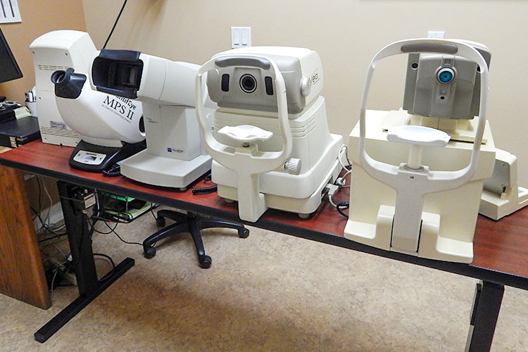
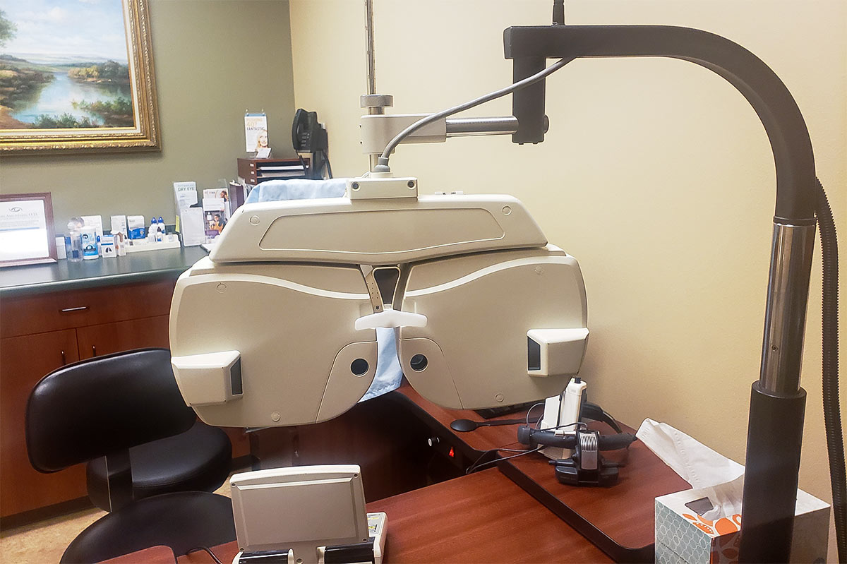
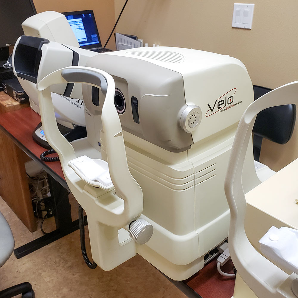
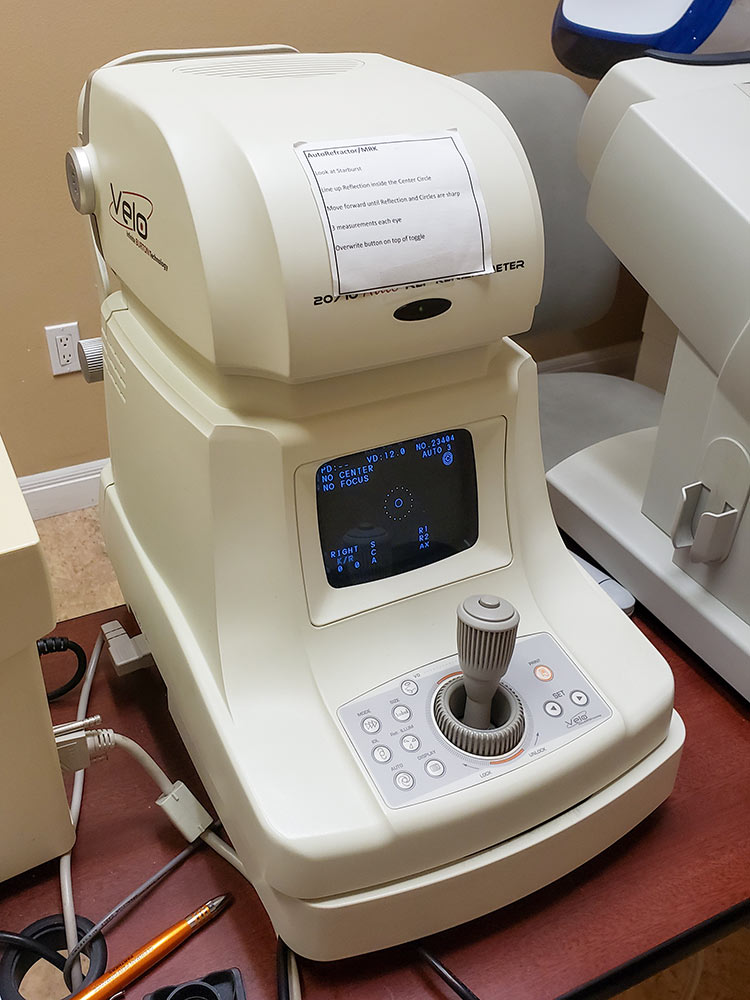
Computerized Vision Testing: Our office utilizes advanced computer assisted instruments to test all aspects of the optics of your eyes including higher order aberrations to determine how to maximize your sight. Included in our vision testing equipment are computerized refraction and measuring equipment to assist us in getting more accurate findings.
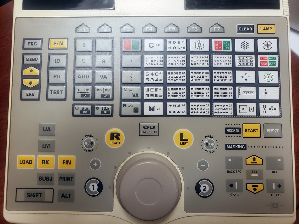
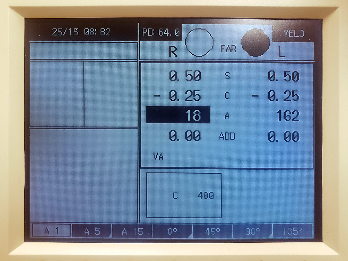
Computerized Refraction: Our testing equipment for glasses allows us to refine our findings in one degree increments while traditional testing is done in 5 degree steps. That is significantly more accurate. The entire process is controlled from a keypad allowing for more controlled testing
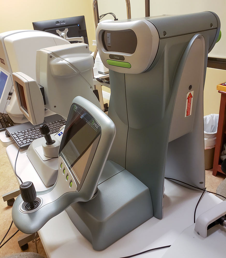
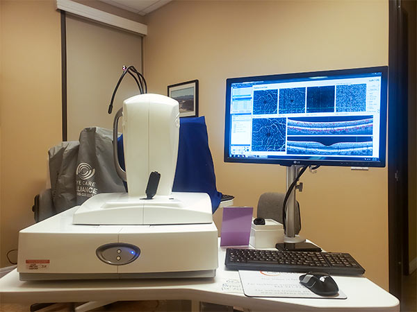
Optical Coherence Tomography (OCT) is a state-of-the-art diagnostic test that allow us to visualize and measurement of structures in the eye including the retina, optic nerve, cornea and iris using very safe visible light. Similar in diagnostic ability to MRI for general body conditions, the images received from the OCT help us diagnose and manage eye conditions such as glaucoma, macular degeneration, and diabetic retinopathy OCT angiography allows us to see vascular changes in the retina to pinpoint the causes of sight loss for early intervention. Below are some examples of testing results from the instrument.
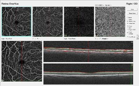
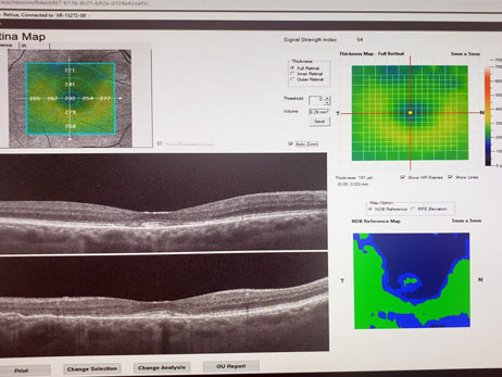
Retina OCT detects structural abnormalities in the center of the retina.
Retinal Imaging
The Optos Daytona captures high resolution 200 degree wide angle views of the retina utilizing low intensity scanning laser technology. We use this instrument to diagnose and manage conditions of the retina. This technology decreases the need and frequency we have to dilate the eyes.


Threshold Visual Field Testing: We have chosen to use the Octopus 600 technology, which originated in Switzerland, because of its precision and speed. This makes testing most accurate and comfortable for our patients. This test is most useful in detecting and treating glaucoma and managing and understanding neurological conditions that affect the eyes.


The detailed information the Octopus 600 provides is invaluable in managing many eye conditions.
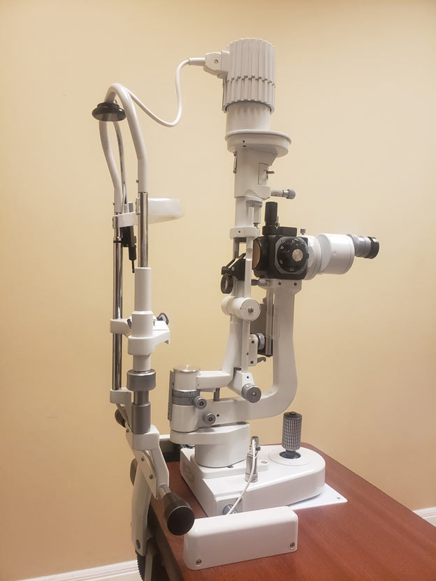
Slit Lamp Biomicroscope: We utilize this instrument daily to examine the eye in 3-D under very high magnification. We are able to diagnose subtle conditions that affect the well-being of your eyes and your precious sight with this instrument.
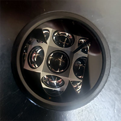
Gonioscopy uses a mirrored instrument to examine the internal fluid drainage structures of the eye which are vital to maintain proper fluid pressure in the eye.
Visually Evoked Potential & Electroretinogram: This technology allows us to assess the health of the retina and the neurological pathway to the brain. Having this information allows us to assess the earliest damage to the eyes from conditions like diabetic retinopathy, glaucoma, and use of Plaquenil. We are able to diagnose conditions like glaucoma as much as eight years before we can detect sight loss.
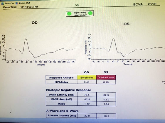
Photopic Negative response test for earliest detection of glaucoma.
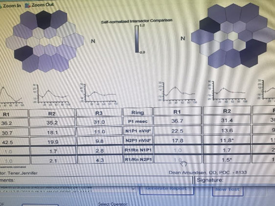
Multifocal Electroretinogram for early detection of Plaquenil retinal toxicity.
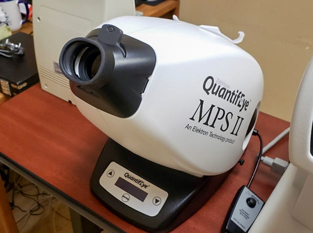
Macular Pigment Optical Density: This technology allows us to measure the amount of macular pigment in the center of the retina called the Macula. The macula allows us to see detail in order to read, recognize faces, and see road signs. Having adequate amounts for macular pigment protects us from damage in the form of macular degeneration. We can help you raise your macular pigment levels.
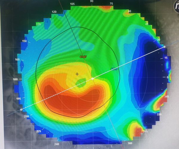
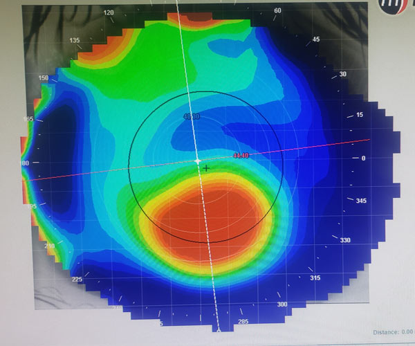
Corneal topography: This instrument measures subtle contour changes in the cornea to help precise fitting of contact lenses and monitor the progression of corneal conditions. Below are some examples of topo maps of the cornea.
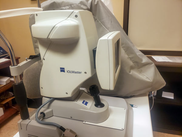
A Scan Ultrasound: We now know that having eyes that are too large can increase your risk of eye disease as you age. The A scan allows us to measure the size of the eye and offer treatments for children to prevent them from having sight problems as adults.
Computerized Patient Records
We maintain computerized patient records, accounting records, and laboratory orders to assure the highest degree of accuracy. Our software allows for you to access your records online from any location. We use federally approved software that is updated regularly as requirements change. Your privacy is very important to us; we conform to HIPAA standards. Our commitment is to provide the highest quality of care for you, your family, and friends. Our computerized patient records meet the stringent government standards regarding privacy. We also implement the latest privacy requirements.

Schedule a Myopia Consultation
We’ll walk you through your options and build a plan tailored specifically to your child.
Call (805) 482-1136 or use our contact form to schedule an appointment with us.